Fast Processing
Our system records images at an impressive 8 seconds per well (specific setting), with up to 96 fields per run. Get results faster than ever before!
seconds recording time per well at the quickest configuration
slides capacity
wells can be processed in one run
traceability
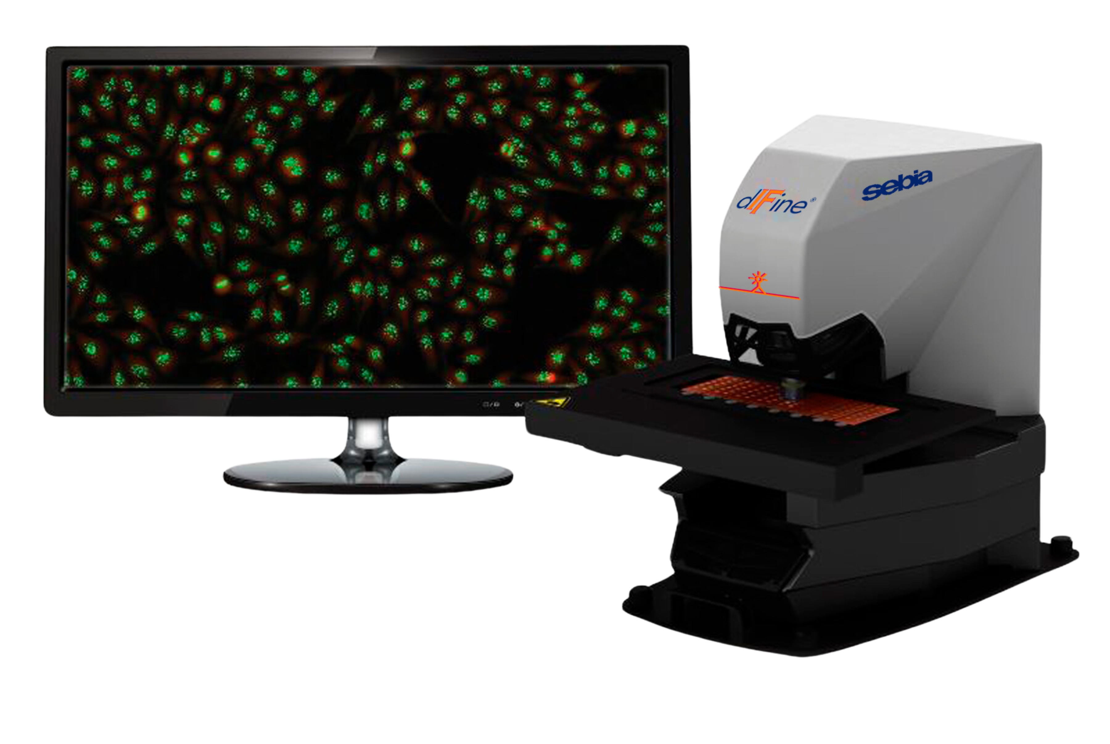
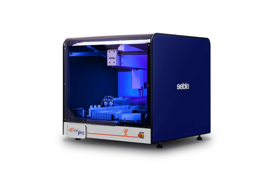
The dIFine® automated microscope is a fully automated IFA microscope, which improves the laboratory workflow significantly. With the small footprint of just 40 x 47cm², it can be accommodated in compact laboratory spaces. The high throughput of 8 seconds per well¹ and the capacity of up to 96 fields per run ensure a long walk away time combined with a high performance. The automated image acquisition, analysis, display and storage streamlines the everyday workflow.
The built-in classifier can identify and suggest different ANA patterns by means of the most advanced artificial intelligence (Homogeneous, Nucleolar, Centromere, Speckled, Cytoplasmic (Ribosomal), Cytoplasmic (Mitochondrial), Nuclear Dots, Nuclear Membrane). Negative samples can be released in batches, so not every single negative picture needs to be evaluated. During the result interpretation also the integrated image atlas ensures easy validations with a possible side-by-side reference.
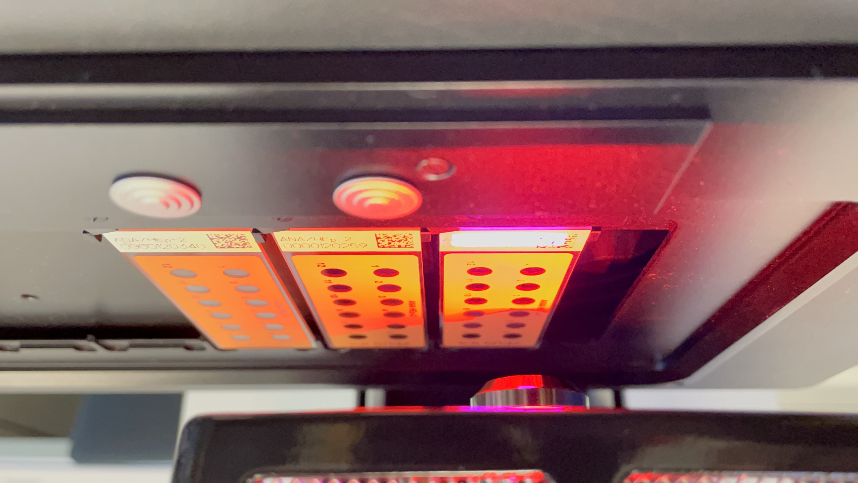
All slides are equipped with a 2D barcode, which identifies the slide, but also includes information about Lot Number and expiration date for a full traceability all the way to the LIS system². The high-quality optics within the microscope provide vibrant colors and the long lasting LED illumination together with the state-of-the-art camera create brilliant pictures for an easy interpretation.
¹ Specific setting
² Compatible LIS required
The dIFine® automated microscope combines advanced AI-based pattern recognition, brilliant optics, and fully automated IFA imaging transforming complex IFA interpretation into a streamlined, reproducible workflow.³
³ Valid if dIFine® automated microscope is used in combination with dIFine® ANA HEp-2 Classifier, dIFine® DNA Classifier, dIFine® ANCA Classifier.
Our system records images at an impressive 8 seconds per well (specific setting), with up to 96 fields per run. Get results faster than ever before!
Validate results confidently. The integrated Image Atlas, aligned with ICAP nomenclature, provides side-by-side references for easy validation.
Harness the power of artificial intelligence. Our built-in classifier recognizes various ANA patterns, including Homogeneous, Nucleolar, Centromere, Speckled, Cytoplasmic (Ribosomal), Cytoplasmic (Mitochondrial), Nuclear Dots, and Nuclear Membrane — providing diagnostic support and streamlining your decision-making process.³
Save time by releasing negative samples in batches. Our system optimizes your workflow, allowing you to focus on what truly matters.³
Never lose track of your slides. The 2D barcode identifies each slide, ensuring full traceability. Plus, data is seamlessly transferred to your LIS system (compatible LIS required).
Experience brilliant images with our high-quality microscope optics. See details like never before.
HEp-2 automatic pattern recognition with suggestion for dIFine® automated microscope used in combination with dIFine® ANA HEp-2 Classifier:
No content available.
Authentically clear!
High Throughput - Smart Automation.
Small Footprint - Big Impact.
Any assay, any sample, any time!
Flash Forward to Accurate Results!
This section contains information intended for wide distribution and may therefore contain product details or information that is not available or valid in your country. Please contact your local Sebia representative. Information intended for healthcare professionals. Carefully read the instructions in the reagent package inserts and instrument manuals.